
Case Study: Successful Removal of Multiple Kidney Stones Using RIRS
Kidney stones are a common yet painful condition that can significantly impact a person’s quality of life. In this case study, we present a 100% real example of a patient who was diagnosed with multiple kidney stones and successfully treated using Retrograde Intrarenal Surgery (RIRS)—a modern, minimally invasive technique.
Patient Diagnosis Based on NCCT KUB Scan
A Non-Contrast Computed Tomography (NCCT) scan of the Kidneys, Ureters, and Bladder (KUB) region was performed. The scan revealed the following findings:
- Both kidneys showed parenchymal thinning and cortical scarring, indicating long-standing stress or infection.
- Right kidney: Measured 8.7 x 4.9 cm with 8–9 non-obstructive calculi in the lower pole. The largest stone was about 1.5 x 1.2 cm.
- Left kidney: Measured 9.2 x 4.6 cm with 7–8 non-obstructive calculi in various regions and a large stone (1.5 x 1.7 cm) in the lower pole.
- A DJ stent was seen in the left kidney, placed earlier to aid in drainage.
- An obstructive stone in the left upper ureter (1.1 x 0.9 cm) was causing mild hydroureter and grade I/II hydronephrosis, a condition where the kidney swells due to urine build-up.
Thankfully, no signs of kidney swelling on the right side or other organ abnormalities were observed. The adrenal glands, urinary bladder, and surrounding fat planes appeared normal.
Treatment Approach – RIRS Procedure
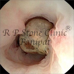
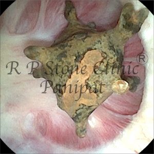
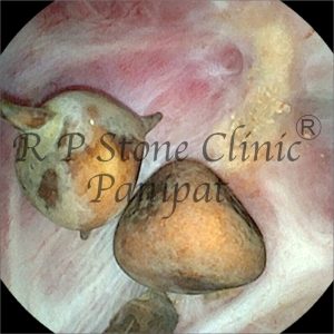
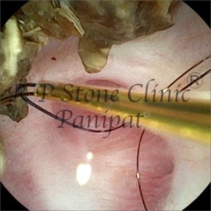
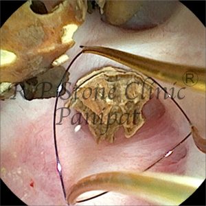
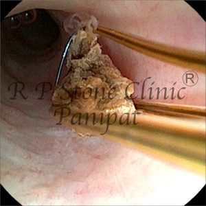
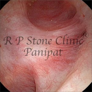
Due to the multiple and varying locations of the stones, our medical team opted for RIRS (Retrograde Intrarenal Surgery). This advanced, scar-free endoscopic procedure is performed using a flexible ureteroscope that passes through the natural urinary tract to access the kidney directly.
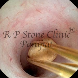
With the help of laser lithotripsy, all accessible stones were broken down and removed in a single or staged procedure. The DJ stent already in place supported post-operative drainage and reduced the risk of complications.
Outcome and Recovery
The patient responded well to the treatment. The stones were successfully cleared without the need for any open surgery or large incisions. Pain reduced significantly within days, and renal function showed signs of improvement. A follow-up imaging and stent removal were scheduled as per protocol.
Key Learnings from This Case
- Early diagnosis using NCCT KUB is crucial in evaluating kidney stone size, position, and severity.
- Multiple stones can still be removed effectively using RIRS without open surgery.
- DJ stenting plays a supportive role in managing obstructive stones and post-operative recovery.
- Minimally invasive techniques reduce hospital stay, pain, and recovery time.
Leave a Reply