

Case Study: Mobile Kidney Stone Causing Occasional Upper Ureter Obstruction
Kidney stones can behave in unpredictable ways. While some remain fixed in one location, others are mobile—moving between the kidney and ureter. These moving stones can intermittently block urine flow, causing fluctuating symptoms and imaging findings.
This case focuses on a patient with right kidney stone, where the right kidney stone’s occasional presence in the upper ureter is believed to be the cause of a dilated ureter.


Key NCCT KUB Findings
Right Kidney:
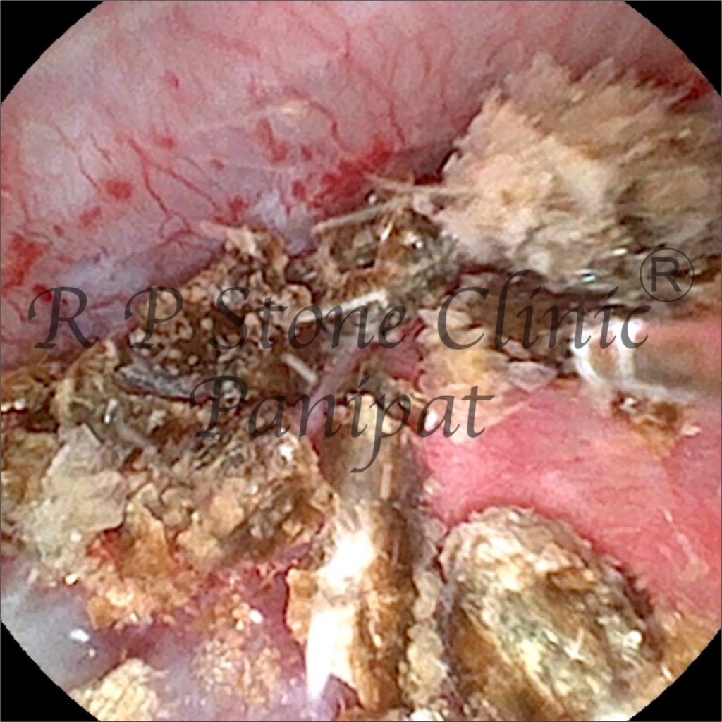
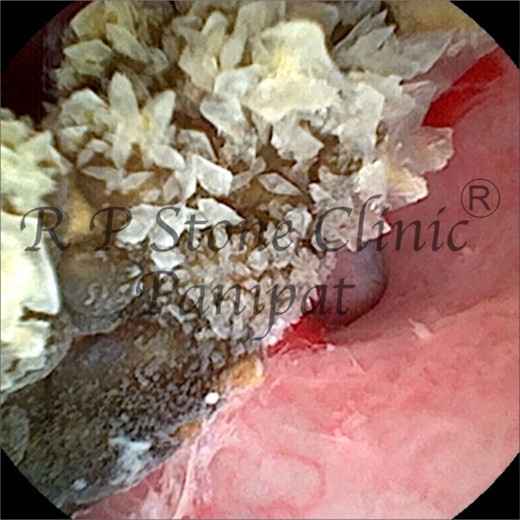
No fixed ureteric stone at the time of scan, suggesting the dilation is due to a stone that occasionally moves into the upper ureter. A calculus of 13 mm x 6.4 mm was seen in the lower pole calyx.
- Ureter dilated in the upper third
- Mild fat stranding near the renal pelvis
- No fixed ureteric stone at the time of scan, suggesting the dilation is due to a stone that occasionally moves into the upper ureter
Interpretation & Clinical Insight
🔹 Why the Ureter is Dilated
In this case, the right ureter’s dilation is due to:
- A mobile stone that occasionally lodges in the upper ureter
- Temporary obstruction when the stone is in the ureter
- Relief of blockage when the stone moves back into the kidney


Treatment Considerations
Possible treatment options:
- RIRS (Retrograde Intrarenal Surgery) is the ideal treatment for such stones.
Preventing Recurrence
- Drink plenty of fluids
- Avoid Non-Vegeterian diet
- Reduce salt and oxalate-rich foods (e.g., spinach, nuts, chocolate)
- Maintain a balanced calcium intake
- Regular imaging follow-ups if prone to recurrent stones
Conclusion
This case highlights the challenges of a mobile kidney stone that can intermittently obstruct the upper ureter.
Although no fixed ureteric stone was found on the scan, the ureteral dilation reflects past or occasional blockages caused by the stone’s movement.
Leave a Reply