
Kidney stones can range from tiny grains to large obstructions that severely affect kidney function. In this case, we highlight a patient who came to our clinic with left-sided abdominal pain, and a CT Urography helped reveal a major finding.
Diagnosis Through CT Urography
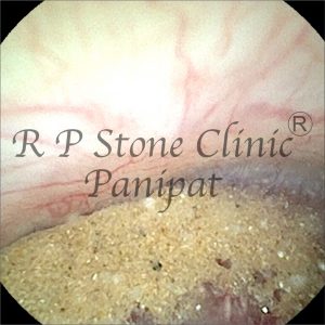
CT Urography is a powerful diagnostic tool that helps visualize the kidneys, ureters, and bladder in great detail using contrast imaging. The scan in this case revealed the following:
Findings:
Right Kidney:
- Normal size: 9.6 x 4.7 cm
- Normal position, outline, and contrast function
- A small non-obstructive stone (2–3 mm) seen in the middle pole
- No swelling or inflammation detected
- Ureter appeared normal
Left Kidney:
- Enlarged: 10.7 x 5.4 cm
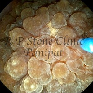
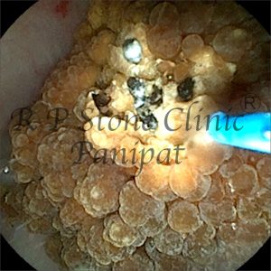
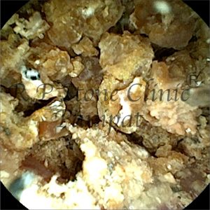
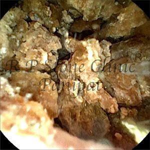
- A large obstructive stone measuring 18 x 17 x 20 mm was found in the renal pelvis, extending into the lower pole calyx
- Moderate hydronephrosis (swelling due to urine buildup) was visible
- Thickening of the nearby lining (urothelium) and para-pelvic fat stranding indicated significant irritation or inflammation
- Ureter showed normal shape and course
Urinary Bladder:
- Normal shape and size
- No abnormal thickening or defects found
Clinical Interpretation
This case clearly shows:
- A tiny stone on the right side, which currently does not require active intervention.
- A large obstructive stone on the left, causing moderate hydronephrosis and inflammatory changes—this requires urgent treatment to prevent kidney damage.
Treatment Plan & Approach
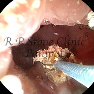
The patient was advised endoscopic removal through RIRS (Retrograde Intrarenal Surgery). This technique allows for:
- Non-invasive stone retrieval
- No external cuts or scars
- Faster recovery time
- Minimal hospital stay
Using flexible ureteroscopy and laser lithotripsy, the large stone can be broken into small fragments and safely removed. Depending on size and shape, DJ stenting may also be used temporarily to relieve the obstruction and allow healing.
Conclusion & Takeaway
This case emphasizes the importance of early detection and imaging in kidney stone cases. CT Urography not only helped identify the size and position of the stones but also guided the correct treatment decision. Left untreated, such obstructive stones can lead to kidney damage, infections, and long-term complications.
Thanks to timely diagnosis and modern endoscopic options like RIRS, patients can now undergo pain-free, scar-free stone removal with excellent outcomes.
Leave a Reply